What Does a B Scan Ocular Ultrasound Capture and Why is it Necessary?
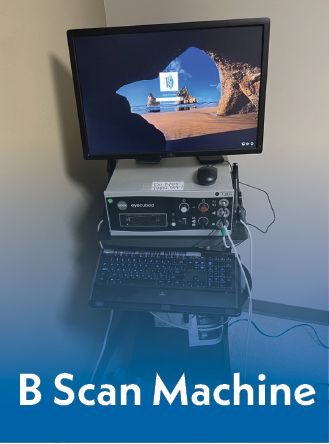
Going to the doctor can be a stressful event for the patient, and over the past few months, we have tried to ease to your worries by sharing information about some of the common testing performed in our office. We are on our last series of blog posts explaining possible imaging that might occur while you are at the office and why. During the exam there may be additional testing ordered, in addition to the physical exam performed by the doctor. This month we are explaining B Scan Ocular Ultrasounds.
What is a B Scan Ocular Ultrasound?
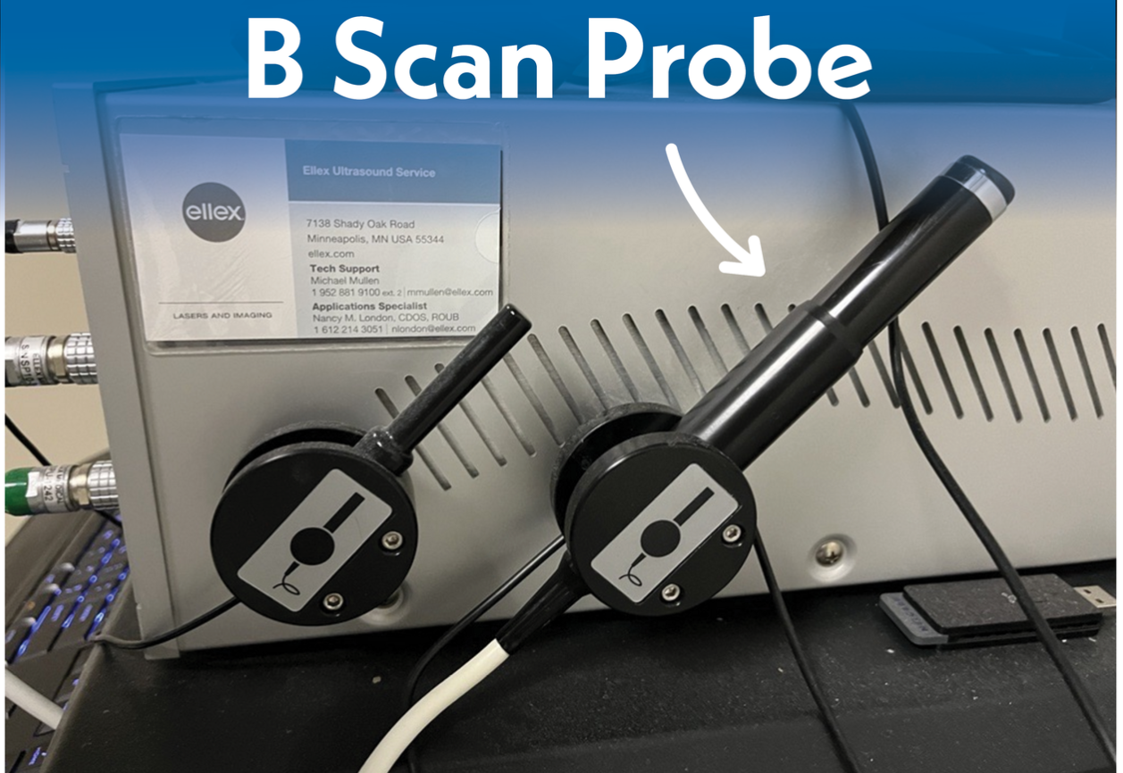
A B-Scan Ocular Ultrasound is used to illustrate a view of your eye by using echoes to produce a bright, detailed image. The ultrasound is a very simple procedure performed to capture a detailed photo of the posterior(the back) portion of your eye. This test may be performed when the view to the back of the eye is obscured or to measure and monitor growth of a lesion. The ultrasound will be performed by our physicians with the assistance of a technician. The doctor will put a lubricant on the probe and ask you to close your eye. This test typically is done on the top of your eyelid. This is not a painful procedure, but you will feel slight pressure on your lid as the doctor takes pictures of different locations of interest. During the scan the doctor will ask you to look in different areas to make sure he captures a detailed photo of the territory that needs to be obtained.
When is a B Scan Ocular Ultrasound necessary?
A B Scan Ocular Ultrasound can be used to discover a wide range of pathological anatomy. A doctor will order a b scan to be performed for multiple reasons. Some of these reasons for a b scan would include retinal detachment, vitreous hemorrhage, dense cataract and tumors. A vitreous hemorrhage or dense cataract for example, will obscure the view for the doctor. This test will allow the doctor to have a better view of the posterior portion of your eye to evaluate the area of concern. A b scan will allow the doctor to view the back of the eye to make sure the patient does not have a retinal detachment or other abnormal pathologies.
Things that may require a B Scan Ocular Ultrasound:
- Retinal Detachment
- Vitreous Hemorrhage (obscures the view of the posterior portion of your eye requiring an ultrasound to be done)
- Dense Cataract (this would also obscure view of the posterior portion of your eye to require an ultrasound)
- Choroidal Melanoma
- Retinoblastoma
- Ocular Trauma
What does a retinal detachment look like on a B Scan Ultrasound Photo?
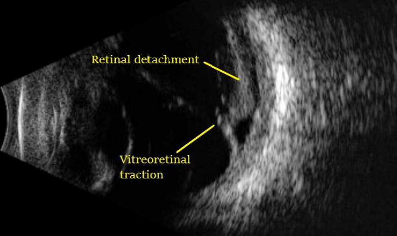
Courtesy of Google Images
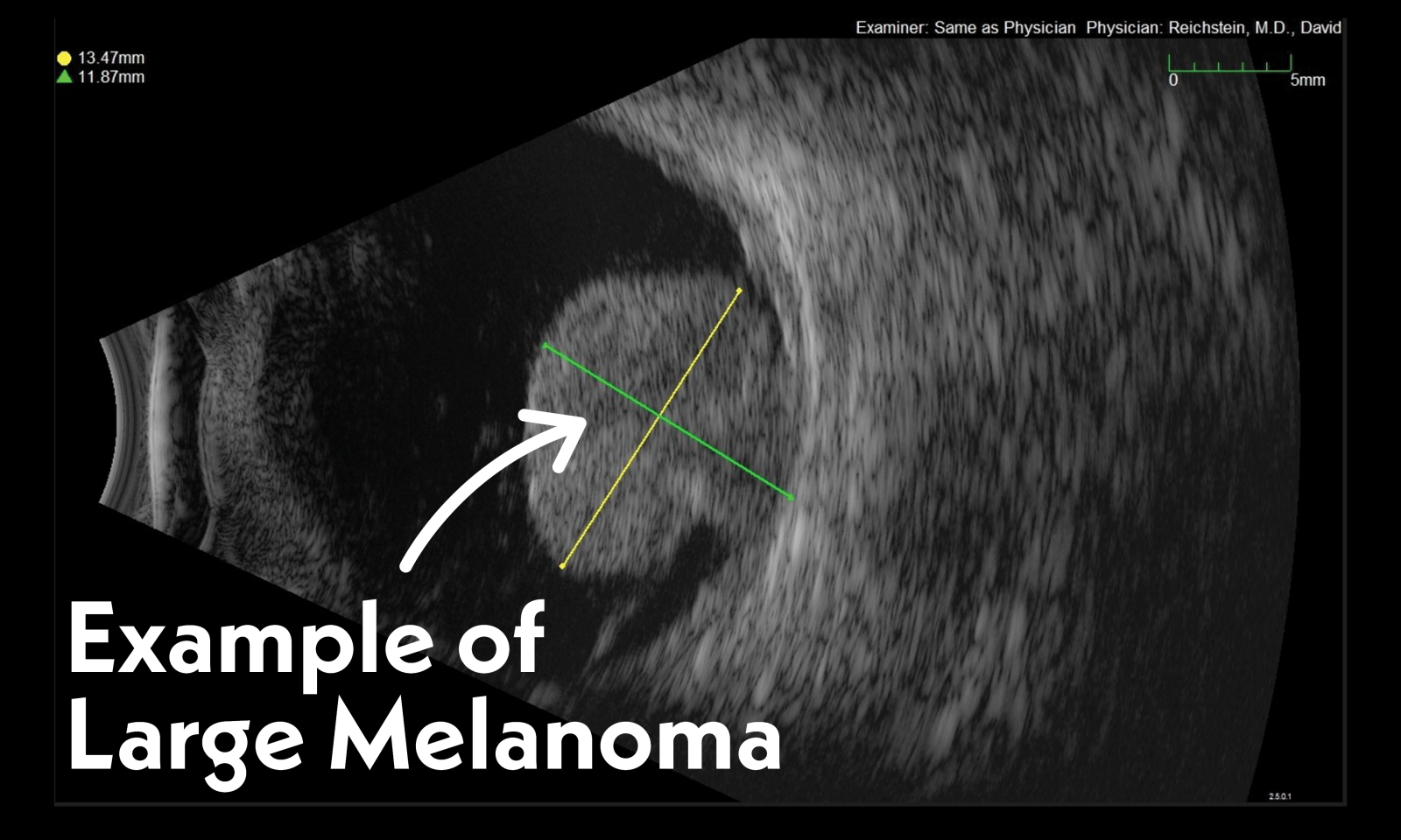
If a tumor has been diagnosed in regular exam, a b scan will be ordered. The ultrasound picture will be able to obtain important measurements including visualization of the tumor, location, size, shape and borders. These measurements are critical.
What does a tumor look like on a B Scan Ultrasound Photo?
What happens next if I am diagnosed with a Choroidal Melanoma?
We have our own oncology team here at TN Retina. Our team is considered one of the largest and most active Ocular Oncology services in the country. Dr. David Reichstein is the director of our Ocular Oncology Service. He is both a vitreoretinal surgeon and an ocular oncologist. He serves along with oncology coordinators Anderson Brock and Holly Lamb to provide exemplary service to the patient. The oncology team will put together the best treatment plan for your specific diagnosis and be alongside of you.
Follow this link to learn more about our oncology team Subspecialty Care > Ocular Oncology (tnretina.com)
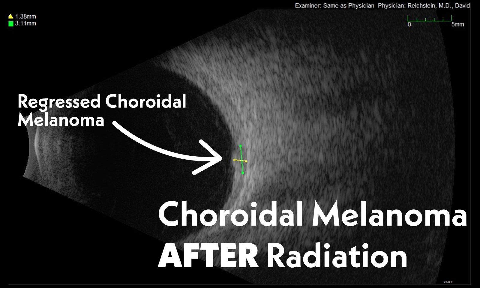
The pictures below represent an actual patient of Dr Reichstein's who was diagnosed with a Choroidal Melanoma. The recommendation for treatment was that the patient proceed with Plaque Brachytherapy. At Tennessee Retina, we perform plaque brachytherapy regularly with excellent rates of tumor control and globe salvage (the ability to keep the eye). Brachytherapy is a broad term used to define the application of radiation directly to a particular body part or tissue. This patient elected to proceed with Radiation therapy. The patient was then scheduled to see a Radiation Oncologist following the diagnosis. The Radiation Oncologist will design a plaque for the individual patient. This plaque will be placed on the melanoma during the surgery performed by Dr Reichstein.
Patient A-Before Tx

Patient A-After Tx
The doctor will examine your retina by looking into your dilated eyes in the exam room, but these tests are very beneficial for the doctor to help confirm the diagnosis and monitor progress if you're undergoing treatment. We hope this series of articles continues to be helpful for you to better understand b scan ocular ultrasounds, OCTs, Fundus photography and fluorescein angiograms and why they may be necessary during your visit with us or your other eye doctors.Coming up next month we will discuss common questions asked by patients in our office, stay tuned!

