What Does a Fluorescein Angiogram Capture and Why is it Necessary?
What does a Fluorescein Angiogram capture and why is it necessary?
Going to the doctor can be a stressful event for the patient. We are continuing our series of blog posts explaining things that might happen while you are at the office and why. During an exam there may be additional testing ordered, in addition to the physical exam performed by the doctor. This month we are explaining Fluorescein Angiography.
What is a Fluorescein Angiogram?
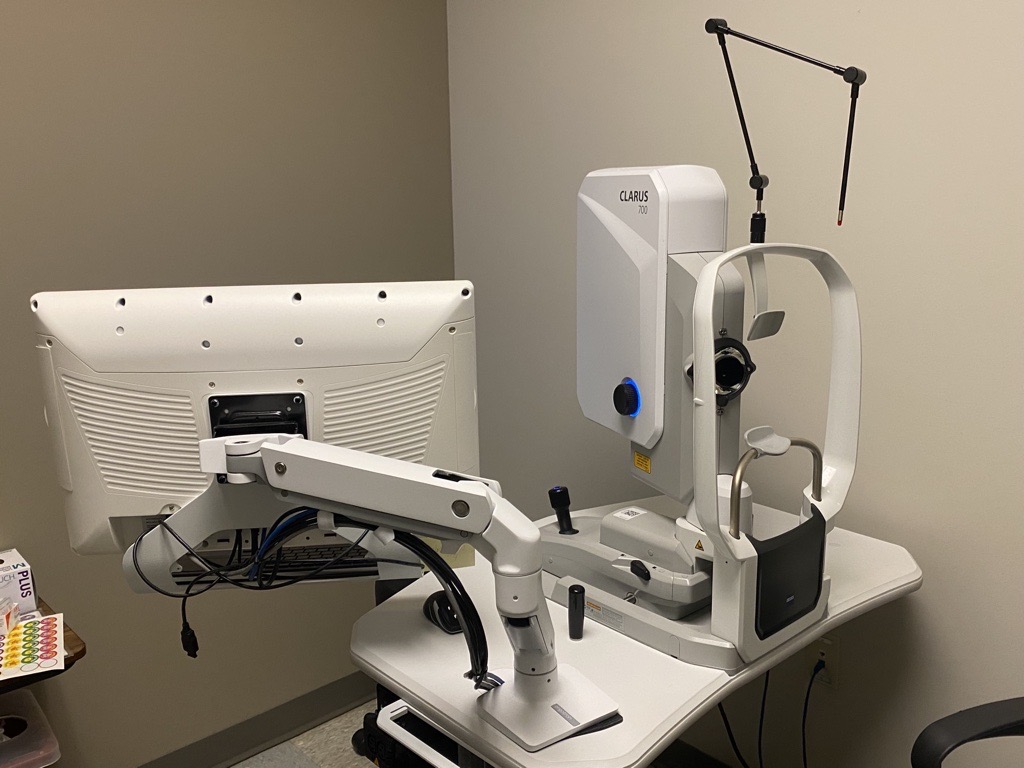
A fluorescein angiogram (FA) is a photo that captures images of the blood flow in the back of your eye. This photo will often be taken on the same camera the photographers use to take a fundus photo (discussed in last month's blog post). You will be asked by one of our photographers to place your chin in the chin rest and place your forehead on the top head rest. There are multiple types of cameras that may be used, but most of them produce a very bright flash, which can be challenging. The blood flow is examined by injecting a small amount of fluorescent dye into your hand or arm. This dye injected into your arm travels through the bloodstream and will highlight (fluoresce) in the blood vessels in the back of your eye due to special filters used in the camera. Through the series of photos taken, your physician can evaluate the blood flow to the retina, including slow blood flow, blockages, or abnormal leakage that may be occurring. The test usually takes 5-10 minutes to perform, and while the bright lights can be a little challenging, the results are very important in assessing the condition of your retina.

When is a fluorescein angiogram necessary?
Fluorescein angiography can help identify a lot of different eye pathologies. Some of these may include diabetic retinopathy, macular degeneration, retinal vein occlusion and vitreous hemorrhages. When any of these diagnoses are suspected by the doctor, this test will likely be ordered. The image will give the doctor a detailed look of the back of your eye and possible areas of concern. This image is also a great way to give the patient a visual of what is happening in the eye and help explain the diagnosis and treatment plan. Angiogram photos also serve as documentation in the patient's health record for comparison and coordinating care.
What does a normal and abnormal fluorescein angiogram look like?
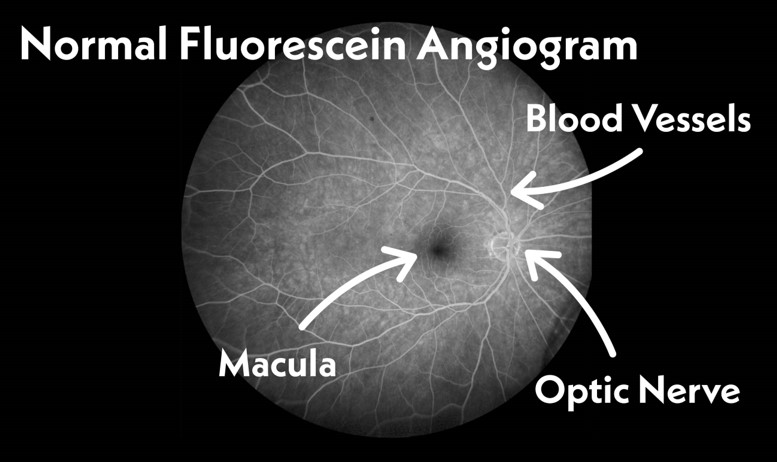
The image below represents a normal fluorescein angiogram (FA) of the retina. This image was captured by one of our very talented TNR photographers. In this image you will be able to identify your optic nerve, macula and vasculature, all with normal blood flow. This is what a normal FA looks like…..

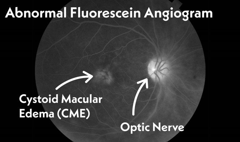
An abnormal fluorescein angiogram could have many presentations. This picture below represents what cystoid macular edema(CME) would look like on a fluorescein angiogram. The CME in this photo below is the highlighted white area to the left of the optic nerve. The CME in the back of the eye usually blocks some or all of the vision depending on where the leakage is located and how severe it is.

The doctor will examine your retina by looking into your dilated eyes in the exam room, but these tests are very beneficial for the doctor to help confirm the diagnosis and monitor progress if you're undergoing treatment. We hope this article was helpful for you to better understand fluorescein angiography and why it may be necessary during your visit with us or your other eye doctors. Stay tuned next month, we will be explaining what a B Scan Ocular Ultrasound is and why it may be necessary.

