What Does an OCT Photo Capture and Why is it Necessary?
Going to the doctor can be a stressful event for the patient. Fear and anxiety often occur when worrying about your health and possible diagnosis. To help put an ease to your worries, we are going to explain some of the things that may occur during your visit and why. During an exam there may be additional testing ordered, in addition to the physical exam performed by the doctor. Throughout the next few months, we are going to try to explain what some of these additional tests are and why they may be ordered by your doctor at your appointment. We hope this helps you understand these tests a little more and make your feel more comfortable at your appointment.
What Is An OCT?
Optical Coherence Tomography(OCT) is used regularly to capture high resolution, cross sectional photos of the back of your eye. The OCT uses a noninvasive laser to capture this image. The image will show the layers of the retina and your optic nerve. The OCT also has the capability to measure the thickness of your retina and optic nerve. This image helps to identify abnormalities and helps confirm the diagnosis during an exam. The OCT is often compared to an ultrasound, but with the OCT there is use of light instead of sound. A few diagnoses that can be picked up by an OCT image include the following: diabetic retinopathy, detached retina, age related macular degeneration and macula hole.
What Is Optical Coherence Tomography Angiography?
Optical Coherence Tomography Angiography captures images of the blood vessels in the back of your eye similar in appearance to an x ray image. This image may be ordered if there is any concern of vascular issues. The image will show blood vessels in detail and can help confirm if there is any leakage present. If the test indicates leakage or other issues that may exist, additional pictures may be ordered including fundus photos or a fluorescein angiogram to help confirm the diagnosis. These additional photos will provide more detail to confirm the diagnosis.
When is a OCT necessary?
Glaucoma- an OCT may be ordered to evaluate the optic nerve.
Age-Related Macular Degeneration(AMD)-an OCT may be ordered to evaluate the retina and check for any leakage or abnormalities present associated with AMD.
Diabetes- if you are diabetic an OCT may be ordered to evaluate the retina for any possible leakage or abnormalities associated with diabetes.
Cystoid Macular Edema(CME)-refers to swelling in your macula (edema). If swelling is suspected, an OCT may be performed to confirm the edema.
Macular Pucker or Epiretinal Membrane(ERM)-scar tissue can grow over the surface of your retina causing distorted vision. When the ERM worsens you sometimes have vision loss, and the OCT will evaluate and confirm the severity of the ERM.
These are just a few examples of when an OCT image is necessary.
What does a normal OCT photo look like?

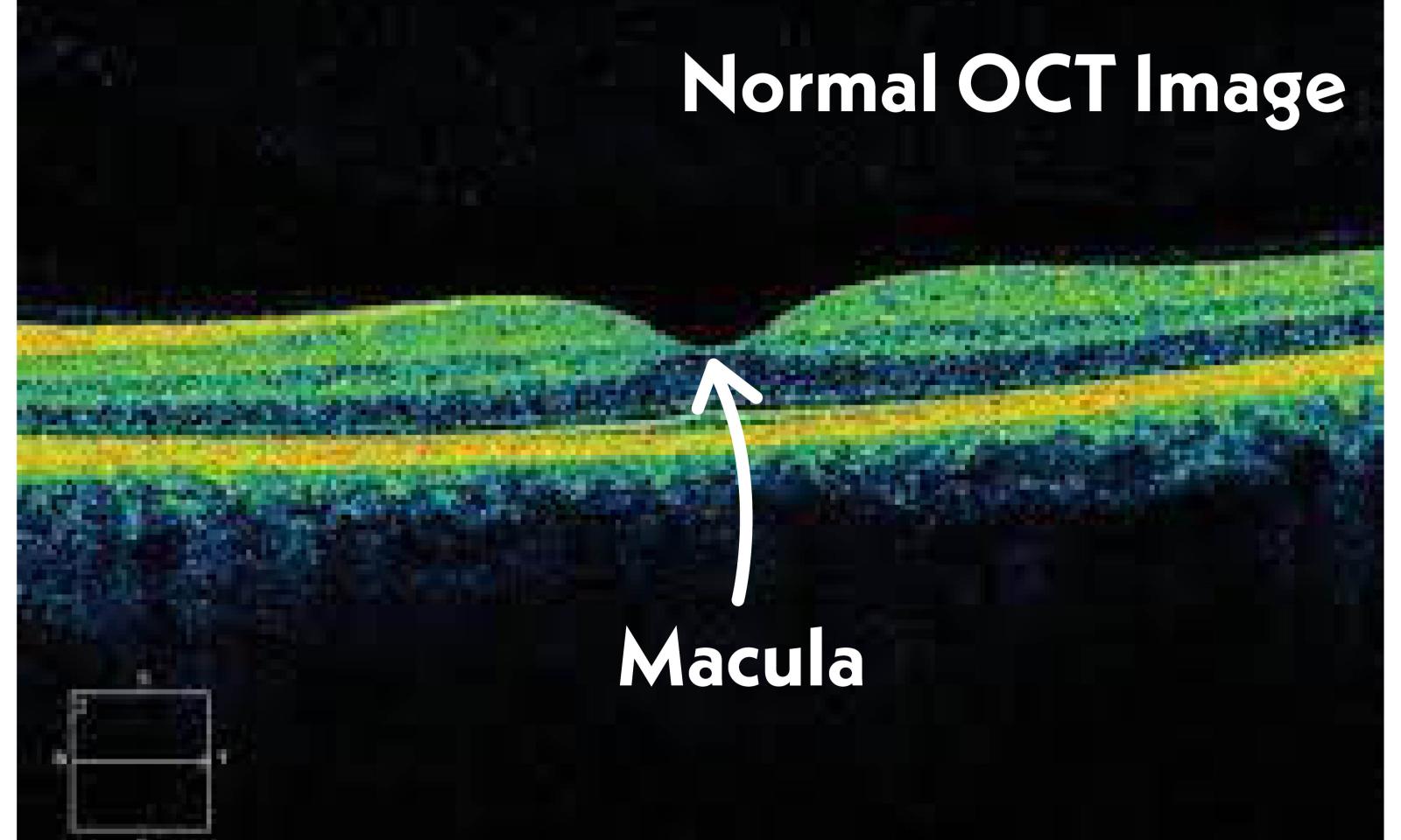
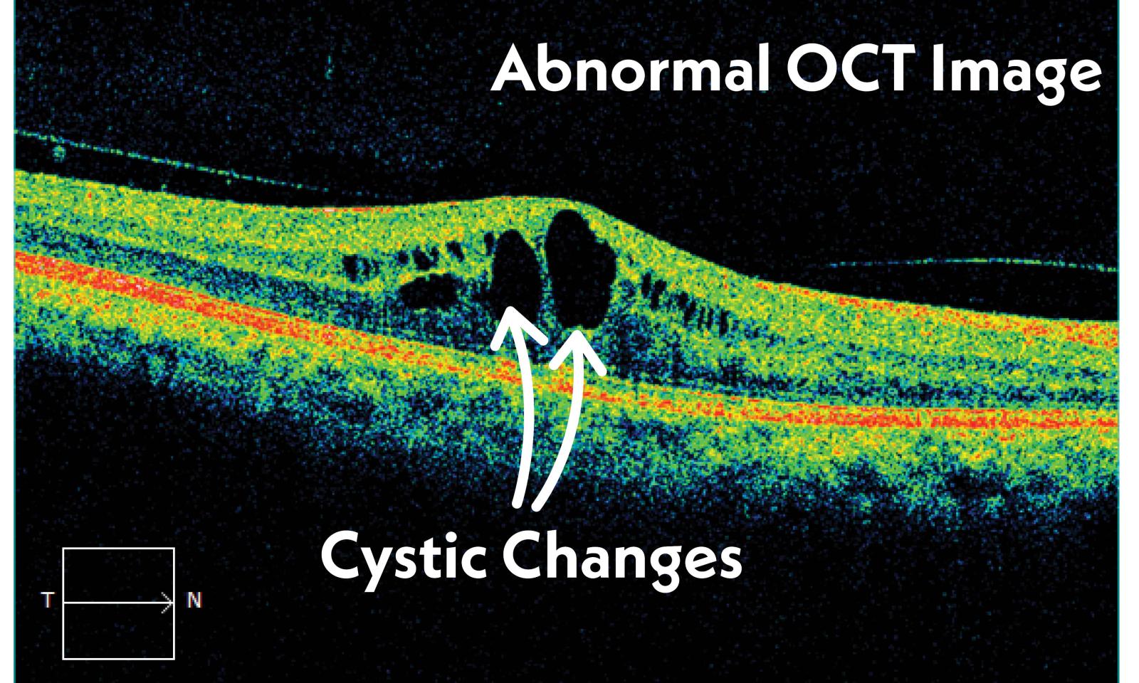
A common question, "is there supposed to be a dip in the middle of my picture?" The answer to this question is YES. The dip shown in the middle of the OCT photo is your macula. The macula is where your central vision comes from. Although the dip is normal there should not be any fluid or cystic changes in your retina. If those changes are present they would look similar to this….

If fluid or cystic changes are noted on your OCT image, additional testing may be ordered including fundus photos and a fluorescein angiogram. These additional photos will look closer into the detail of the blood vessels and other structures in the back of your eyes. The doctor will examine your retina by looking into your dilated eyes in the exam room, but these tests are very beneficial for the doctor to help confirm the diagnosis and monitor progress if you're undergoing treatment. We hope this article was helpful for you to better understand the OCT image and why it may be necessary during your visit with us or your other eye doctors.


